How to conduct an electrocardiogram: ECG best practices
Last Updated on 19 de December de 2023 by Monica Jorge
The electrocardiogram (ECG) stands as a cornerstone in the evaluation of a patient’s cardiac health. Despite its apparent simplicity, the ECG best practices are paramount for an accurate diagnosis. For this reason, in this article we explain how to conduct an eletrocadiogram.
Any type of noise or interference when performing an electrocardiogram can mask serious problems or even make it impossible for the doctor to interpret the test.
It is worth remembering that since some heart diseases are time-sensitive,a poorly conducted ECG could delay crucial results. This can cost the patient’s life, as in the case of a heart attack, for instance.
Throughout this text, we will address some crucial points of performing an ECG correctly, detailing how to conduct the examination, properly attach electrodes to the body, and accurately interpret the results.
ECG best practices: how to conduct an eletrocardiogram ?

For learn how to conduct an eletrocardiogram you have to know the ECG best practices, it is important to know that the electrocardiogram involves understanding the anatomical electrical impulses necessary for the contraction of the myocardium. It provides an electrical evaluation of the patient’s cardiac activity presented graphically.
Through ECGs, various heart conditions and other physiological abnormalities affecting the electrocardiographic recording can be diagnosed, including:
- Old or acute myocardial infarctions (in evolution at that time);
- Inflammation of the heart muscle or membrane lining the heart
- Other cardiovascular complications.
The test can also show consequences in the heart derived from lung diseases like emphysema or pulmonary embolism and some congenital abnormalities.
Read more: Telecardiology: capabilities and features
Correct Placement of Electrodes for Cardiac Monitoring
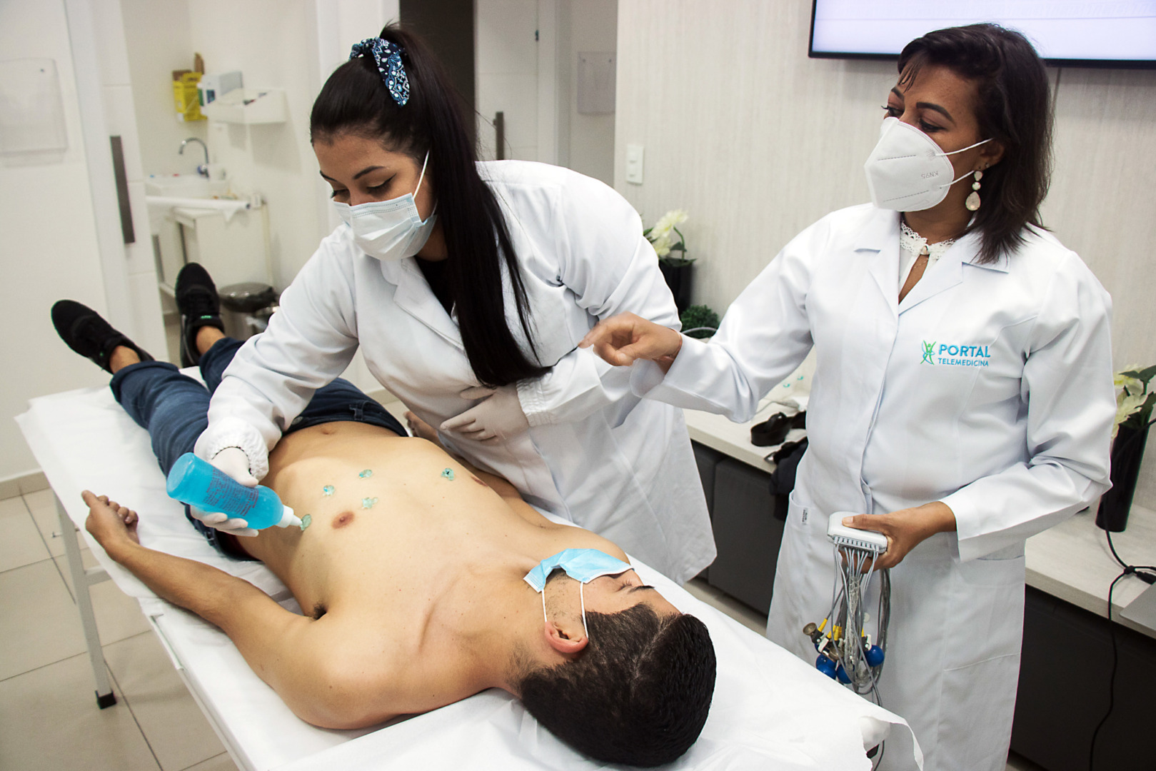
A nursing professional uses an electrocardiograph to conduct the ECG best practices. The apparatus is connected to the patient using Electrodes – small devices that come into contact with clean skin and are fixed with conductive gel in their cups. The gel facilitates the capture of stimuli.
Ten devices, categorized by color, guide precise electrode placement on the patient based on specific criteria.
The patient should lie down, and the electrodes should be placed on the chest, wrists, and ankles. The devices placed on the chest should be in positions V1 to V6, forming the horizontal plane for recording the electrical activity of the heart. The ECG test takes 5 to 15 minutes to complete.
With the test, it is possible to detect the heart rhythm and the number of beats per minute. A normal heart rate ranges from 50 to 90 beats per minute. Rates exceeding 100 or dropping below 40 beats per minute indicate elevated or reduced heart function, respectively.
It’s crucial to note that while nursing professionals can conduct the ECG, only cardiologists can interpret the data and provide diagnoses.
Equipment Used for Electrocardiograms (ECG)
The ECG is performed using an electrocardiogram machine, also known as an electrocardiograph, which analyzes heart frequencies and signs of failure through graphical representation. These machines can be either digital or analog.
High-quality devices improve data interpretation and diagnostic safety, so it is vital to choose models that generate high-resolution images and ensure their regular maintenance.
The electrocardiogram device must be registered by the Brazilian Health Regulatory Agency, Anvisa. If the clinic opts for more updated digital devices, they can easily connect them to telemedicine services, which allow for remote analysis and online reporting.
Read more: Digital health: What is it and what are its benefits?
Common ECG Changes
As explained earlier, the electrocardiogram shows well-defined electrocardiographic patterns through graphs.
When conducting an ECG, specific elements like waves and intervals are crucial for accurate results.
Each registered contraction is represented by an irregular line, which can be broken down into small segments or “waves,” designated by letters, signifying heart chamber activation.
A change in the configuration of these “waves” may indicate the presence of a heart disturbance.
Errors in Conducting an ECG – and How to Avoid Them
Nursing professionalS responsible for performing the electrocardiogram need to be aware of the most common errors that can occur during the exam. These errors hinder correct ECG execution, which makes it impossible for the cardiologist to make an accurate diagnosis.
Among the most common errors, we can mention:
- Leaving the filter off, interferING with graph reading;
- Cable swapping, poor maintenance, or lack of channel: a channel may have interference in the straight wave, and the nursing professional may not notice the error;
- Misplacement or incorrect positioning of electrodes, resulting in inaccurate results
See in the following images how the normal tracing of the exam should be and some examples of deviations caused by examination errors:
Normal tracing:
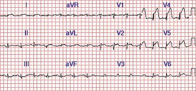
Tracing with Error – 60Hz Filter Off:
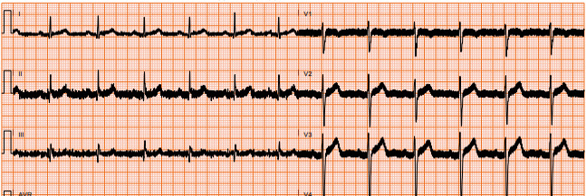
Tracing with Error – lack of Channel (Channel Off):

Telemedicine speeds up ECG reports delivery
Now that you know how to conduct an electrocardiogram, did you know that clinics and hospitals are utilizing telemedicine for ECG reporting and having a renowned cardiologist available 24/7 to interpret the results?
Portal’s tele-report solution has the precision of machine learning algorithms and artificial intelligence for screening exams, prioritizing urgent cases in the queue. Consequently, the company delivers reports in up to 5 minutes for critical situations..
Here’s how it works: Your nursing team conducts the ECG using Portal’s advanced technology, the device sends real-time data to a medical center, then machine learning and artificial intelligence algorithms prioritize exams with alterations, and from there, a qualified specialist provides accurate reporting based on the data.
Utilizing secure remote examinations significantly reduces costs and enhances your team’s productivity and service capacity.
Speak to one of our consultants and find out how easy it is to implement a telemedicine solution in the clinic where you work.

Article translated by Celen Diaz
Graduated in Modern Languages and Business Translation,
with more than 10 years of experience as a Linguist.


